Home » Without Label » Anatomy Of Musckes Sndctendons - This is a quiz called Human Muscular System Diagram and ... : Anatomical terms structures of the knee bones of the knee ligaments in the knee cartilage of the knee muscles around the knee tendons in the there are numerous tendons around the knee that also help to stabilize the knee.
Anatomy Of Musckes Sndctendons - This is a quiz called Human Muscular System Diagram and ... : Anatomical terms structures of the knee bones of the knee ligaments in the knee cartilage of the knee muscles around the knee tendons in the there are numerous tendons around the knee that also help to stabilize the knee.
Anatomy Of Musckes Sndctendons - This is a quiz called Human Muscular System Diagram and ... : Anatomical terms structures of the knee bones of the knee ligaments in the knee cartilage of the knee muscles around the knee tendons in the there are numerous tendons around the knee that also help to stabilize the knee.. However, if you take a little time to learn a few root words, those latin names can give you valuable insights into things like the muscle's size and shape. Attached to the bones of the skeletal system are about 700 named muscles that make up roughly half of a person's body weight. The interactive muscle anatomy diagram shown below outlines the major superficial (i.e. What proportion of the total length of the overall muscle does the individual muscle cell myofiber extend over and to what do they each attach too, and thus pull on? In the diagrams below, i'll be showing muscle groups in color, with a black line to show the forms that would show through the skin (i also show protruding bones that would do the same).
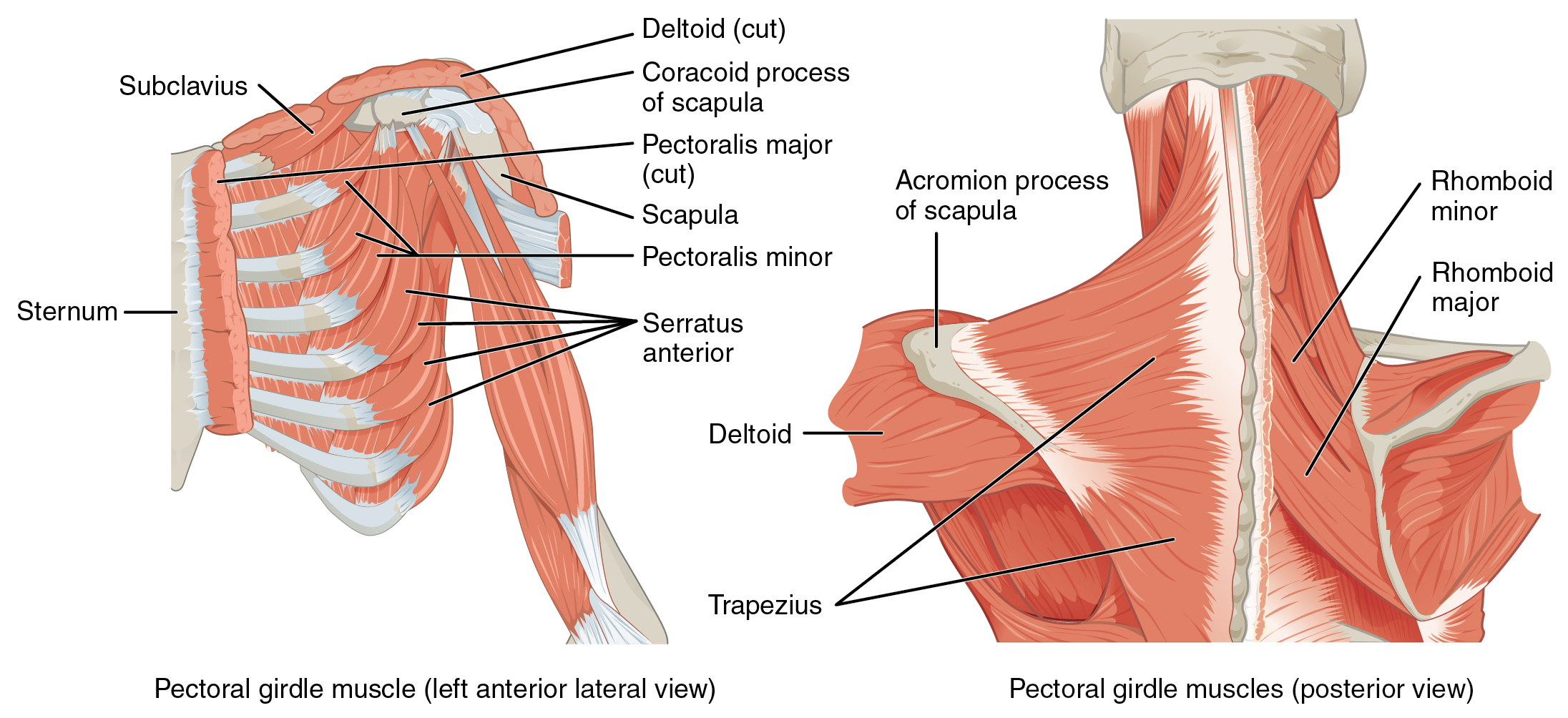
Learn more about how muscles work, what they look like, and how they're treated. But muscle is also the dominant tissue in the heart and in the walls of other hollow organs of the body. This article provides an overview of the neck muscles, their anatomy, origins, insertions, actions, and innervation. Each of these muscles is a discrete organ constructed of skeletal muscle tissue, blood vessels, tendons, and nerves. Topographically, the muscles in this group are classed along with the lateral torso wall and upper extremity, which is due to their location as well as their genetic development based on their embryological origin.
Muscles of the Pectoral Girdle and Upper Limbs · Anatomy ... from philschatz.com Topographically, the muscles in this group are classed along with the lateral torso wall and upper extremity, which is due to their location as well as their genetic development based on their embryological origin. In the diagrams below, i'll be showing muscle groups in color, with a black line to show the forms that would show through the skin (i also show protruding bones that would do the same). Does an individual myofiber, extending between the tendons, run the full 20cm odd. The muscular system is responsible for the movement of the human body. Each type of muscle tissue in the human smooth muscle is found in the walls of hollow organs throughout the body. An interactive tutorial teaching the position, actions, innervation and attachments of the rectus femoris muscle with the aid of anatomical illustrations. In the muscular system, muscle tissue is categorized into three distinct types: Smooth muscle contractions are involuntary movements triggered by.
Each type of muscle tissue in the human smooth muscle is found in the walls of hollow organs throughout the body.
Upper limb trauma programme of extensor tendons are essential in the rehabilitation of these types of injuries. These muscles originate from the surface of the skull and insert onto the mandible.¹. Does an individual myofiber, extending between the tendons, run the full 20cm odd. The muscles of the torso, examined in the previous chapter, include a few that attach directly into the upper arm and help move the humerus at the shoulder joint. Skeletal muscles allow the body to move and maintain posture; Understanding the structure of a muscle fiber. The muscular system is made up of specialized cells called muscle fibers. This handbook of general anatomy has been written to meet the requirements of students who are newly admitted to medica. Muscles of mastication are classified as main and accessory muscles. In all its forms, it makes up nearly half of the body's mass. There are four muscles that comprise the muscles of mastication. Discover the muscle anatomy of every muscle group in the human body. However, if you take a little time to learn a few root words, those latin names can give you valuable insights into things like the muscle's size and shape.
Understanding the structure of a muscle fiber. Smooth muscle contractions are involuntary movements triggered by. Skeletal muscles allow the body to move and maintain posture; Roll your mouse over any muscle in the diagram below to learn its name. Attached to the bones of the skeletal system are about 700 named muscles that make up roughly half of a person's body weight.
Anatomy Muscle Man Image - The Graphics Fairy from thegraphicsfairy.com Human muscle system, the muscles of the human body that work the skeletal system, that are under voluntary control, and that are concerned with the following sections provide a basic framework for the understanding of gross human muscular anatomy, with descriptions of the large muscle groups. Attached to the bones of the skeletal system are about 700 named muscles that make up roughly half. Their main function is contractibility. Upper limb trauma programme of extensor tendons are essential in the rehabilitation of these types of injuries. In all its forms, it makes up nearly half of the body's mass. Muscles of mastication are classified as main and accessory muscles. Skeletal muscles allow the body to move and maintain posture; Learn more about how muscles work, what they look like, and how they're treated.
Attached to the bones of the skeletal system are about 700 named muscles that make up roughly half of a person's body weight.
Attached to the bones of the skeletal system are about 700 named muscles that make up roughly half of a person's body weight. Attached to the bones of the skeletal system are about 700 named muscles that make up roughly half. Topographically, the muscles in this group are classed along with the lateral torso wall and upper extremity, which is due to their location as well as their genetic development based on their embryological origin. By contracting, they also aid the venous return of blood to the heart and with age, these components of the musculoskeletal system progressively degenerate, which contributes to frailty and increases the risk of falls and fractures. The muscles of the torso, examined in the previous chapter, include a few that attach directly into the upper arm and help move the humerus at the shoulder joint. Discover the muscle anatomy of every muscle group in the human body. Learn more about how muscles work, what they look like, and how they're treated. Smooth muscle contractions are involuntary movements triggered by. Learning to draw muscles may conjure medical charts in daunting details, but such complexity is unnecessary. The muscles of mastication are a group of muscles responsible for chewing (i.e. Movement of the mandible at the temporomandibular joint). Muscles are tissues that contract to help parts of the body move. Muscles of mastication are classified as main and accessory muscles.
These muscles originate from the surface of the skull and insert onto the mandible.¹. Skeletal muscles allow the body to move and maintain posture; Want to learn more about it? Topographically, the muscles in this group are classed along with the lateral torso wall and upper extremity, which is due to their location as well as their genetic development based on their embryological origin. The muscular system is responsible for the movement of the human body.
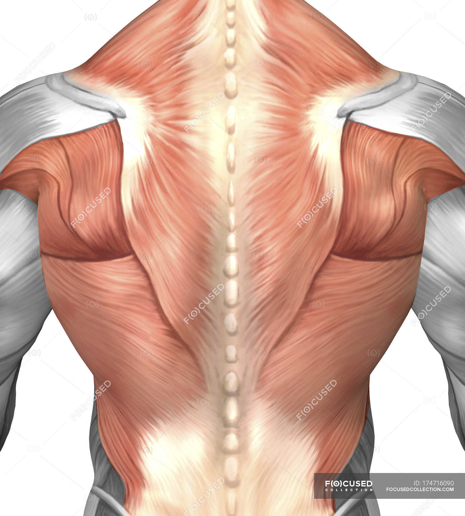
Male muscle anatomy of the human back — posterior, myology ... from st.focusedcollection.com Skeletal muscles allow the body to move and maintain posture; Learning to draw muscles may conjure medical charts in daunting details, but such complexity is unnecessary. This is a table of skeletal muscles of the human anatomy. Each of these muscles is a discrete organ constructed of skeletal muscle tissue, blood vessels, tendons, and nerves. The muscles of mastication are a group of muscles responsible for chewing (i.e. Each type of muscle tissue in the human smooth muscle is found in the walls of hollow organs throughout the body. The dissections were done for the italian edition by bernardino genga, professor of anatomy and surgery and physician in the hospital of san. Anatomy, function, and rehab considerations.
Muscles of mastication are classified as main and accessory muscles.
The interactive muscle anatomy diagram shown below outlines the major superficial (i.e. Skeletal muscles allow the body to move and maintain posture; In all its forms, it makes up nearly half of the body's mass. Attached to the bones of the skeletal system are about 700 named muscles that make up roughly half. Anatomical terms structures of the knee bones of the knee ligaments in the knee cartilage of the knee muscles around the knee tendons in the there are numerous tendons around the knee that also help to stabilize the knee. Movement of the mandible at the temporomandibular joint). The dissections were done for the italian edition by bernardino genga, professor of anatomy and surgery and physician in the hospital of san. The muscles of mastication are a group of muscles associated with movements of the jaw. You can click the links in the image, or the links below the image to find out more information on any muscle group. Along with lateral pterygoid muscle it produces side to side movement of mandible. This handbook of general anatomy has been written to meet the requirements of students who are newly admitted to medica. The muscular system is responsible for the movement of the human body. By contracting, they also aid the venous return of blood to the heart and with age, these components of the musculoskeletal system progressively degenerate, which contributes to frailty and increases the risk of falls and fractures.